
Profiles in Biotech (9/1/23)
Analysis
Axial: https://linktr.ee/axialxyz
Axial partners with great founders and inventors. We invest in early-stage life sciences companies such as Appia Bio, Seranova Bio, Delix Therapeutics, Simcha Therapeutics, among others often when they are no more than an idea. We are fanatical about helping the rare inventor who is compelled to build their own enduring business. If you or someone you know has a great idea or company in life sciences, Axial would be excited to get to know you and possibly invest in your vision and company . We are excited to be in business with you — email us at info@axialvc.com

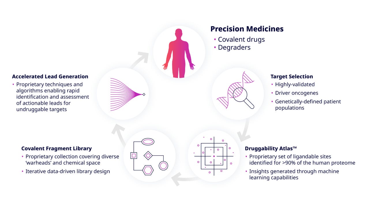
Frontier Medicines develops medicines against disease-causing proteins previously considered undruggable. Founded in 2016, the company's platform is based on a combination of technologies: chemoproteomics, covalent drug discovery & machine learning. Developing a pipeline of precision medicines for cancer along with a collaboration with AbbVie.
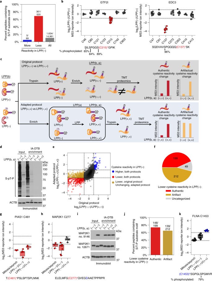
Global profiling of phosphorylation-dependent changes in cysteine reactivity
The paper studies how phosphorylation of proteins affects the reactivity of cysteine residues. Cysteine is an amino acid that is prone to chemical modification, so changes in its reactivity can have a big impact on the function of proteins.
The authors developed a method called TMT-ABPP that can be used to measure the reactivity of cysteine residues on a large scale. They used this method to study the reactivity of cysteine residues in mitotic cells, which are cells that are dividing. Finding that many cysteine residues in mitotic cells undergo phosphorylation-dependent changes in reactivity. Some cysteine residues become more reactive after phosphorylation, while others become less reactive. The authors also found that these changes in reactivity are often localized to specific proteins or regions of proteins. And suggesting that phosphorylation can have a significant impact on the chemical properties of proteins.
https://www.nature.com/articles/s41592-022-01398-2
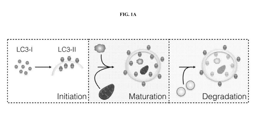
In vitro methods of monitoring autophagy in a human cell using a photoconvertable fluorescent protein linked biomarker
The patent describes a method for monitoring autophagy in a human cell using a photoconvertable fluorescent protein (fcFP) linked to an autophagy biomarker. The fcFP is a protein that can change its fluorescence properties when exposed to light. This allows scientists to track the movement and activity of the autophagy biomarker in the cell.
The specific autophagy biomarker used in the patent is LC3, which is a protein that is involved in the formation of autophagosomes, which are sacs that contain cellular waste. The fcFP is fused to LC3 using a linker segment. This allows the fcFP to follow the movement of LC3 as it is incorporated into autophagosomes.
The method also uses LC3-fcFP fusion protein to measure the induction, maturation, and flux of autophagy in a cell. Induction refers to the process of activating autophagy, maturation refers to the process of forming autophagosomes, and flux refers to the rate at which autophagosomes are being formed and degraded. Providing a new way to monitor autophagy in a cell. And could also be used to develop new drugs that target autophagy.
https://patentimages.storage.googleapis.com/66/a1/c3/266dccf6aaad7b/US10928381.pdf
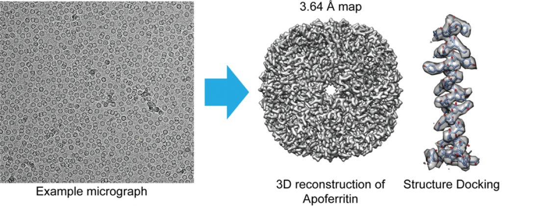
How is protein structure determined by cryo-EM?
Cryo-electron microscopy (cryo-EM) is a technique used to determine the three-dimensional structure of proteins and other biological molecules. In cryo-EM, a sample of the protein is rapidly frozen in a thin layer of liquid ethane, which preserves the molecules in their native state. The frozen sample is then placed on an electron microscope grid and imaged with a beam of electrons.
The electrons interact with the atoms in the protein, creating a diffraction pattern. This pattern is then used to reconstruct a three-dimensional image of the protein. The resolution of the image can be improved by averaging the images of many individual proteins.
The ideal protein size for cryo-EM is between 200 and 1000 kDa. Smaller proteins can be imaged, but the resolution is typically lower. Larger proteins are more difficult to image because they are more likely to aggregate. And has become a powerful tool for structural biology.
It has been used to determine the structures of many proteins that are difficult or impossible to crystallize, including membrane proteins and proteins that are involved in disease:
- Prepare the sample: the protein is purified and then diluted in a solution that will help it to freeze rapidly
- Freeze the sample: flash-frozen in liquid ethane, which preserves the molecules in their native state
- Prepare the grid: frozen sample is applied to a thin carbon film on an electron microscopy grid
- Image the sample: grid is placed in an electron microscope and images are taken of the individual protein molecules
- Process the images: using computer software to reconstruct a three-dimensional image of the protein
Scientist Stories: Shinya Yamanaka, Cell reprogramming and Pioneering Induced Pluripotent Stem Cells
In 2006, Yamanaka discovered that adult somatic cells can be reprogrammed into an embryonic-like pluripotent state by delivering transcription factors