
Profiles in Biotech (8/31/23)
Analysis
Axial: https://linktr.ee/axialxyz
Axial partners with great founders and inventors. We invest in early-stage life sciences companies such as Appia Bio, Seranova Bio, Delix Therapeutics, Simcha Therapeutics, among others often when they are no more than an idea. We are fanatical about helping the rare inventor who is compelled to build their own enduring business. If you or someone you know has a great idea or company in life sciences, Axial would be excited to get to know you and possibly invest in your vision and company . We are excited to be in business with you — email us at info@axialvc.com

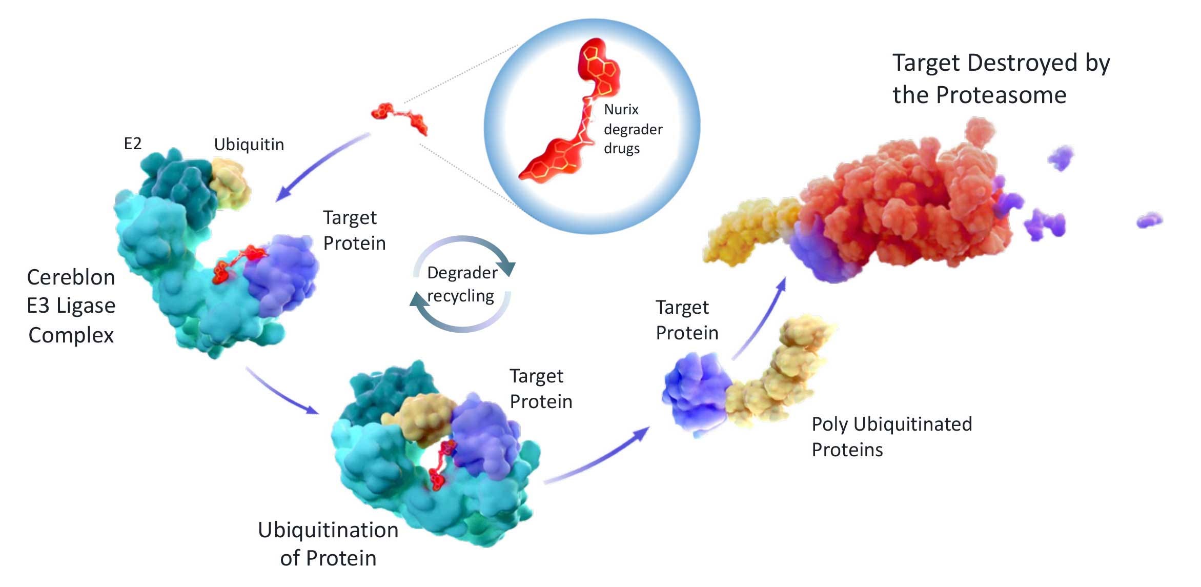
Nurix develops targeted protein degraders (TPD). Small molecule drugs that harness the activity of a class of enzymes called E3 ligases to degrade specific proteins within the cell. That can be used to treat a variety of diseases, including cancer, autoimmune disorders, and neurodegenerative diseases.
Nurix's DELigase platform uses DNA-encoded libraries to screen chemical matter that bind E3 ligases, which are picked for their ability to degrade disease-causing proteins particularly in immune cells. With a lead asset, NX-2127, targeting BTK & IKZF to treat B-cell malignancies.
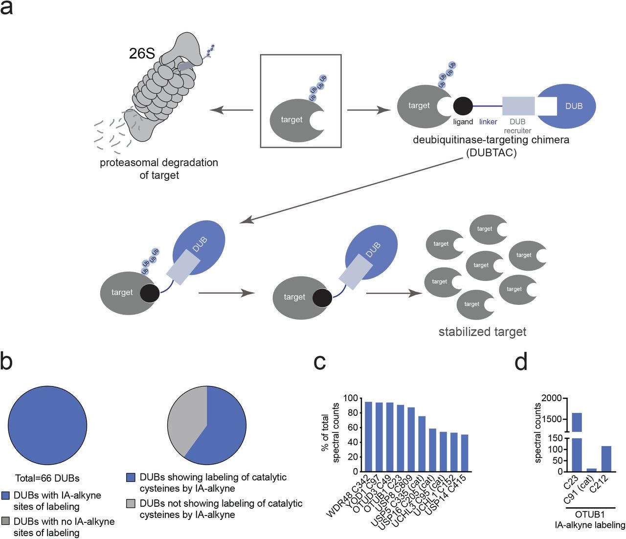
Deubiquitinase-targeting chimeras for targeted protein stabilization
The paper describes an approach to targeting proteins for stabilization, called DUBTACs (deubiquitinase-targeting chimeras). DUBTACs are heterobifunctional molecules that consist of a protein-targeting ligand linked to a DUB recruiter. The DUB recruiter binds to an allosteric site on the DUB, which recruits the DUB to the target protein. This leads to the deubiquitination and stabilization of the target protein.
Using chemoproteomic approaches to identify DUBs that have potential ligandable allosteric sites, the authors found that many DUBs have cysteine residues that are modified by reactivity-based probes, but are not located in the catalytic site. These cysteine residues are therefore potential targets for DUB recruiters.
They then designed and synthesized a DUB recruiter that targets a non-catalytic cysteine (C23) in the K48-ubiquitin-specific deubiquitinase OTUB1. They showed that this DUB recruiter could be linked to a protein-targeting ligand to form a DUBTAC. The DUBTAC was able to stabilize the CFTR protein in cells, which is a protein that is mutated in cystic fibrosis. Concluding that DUBTACs are a promising new approach to targeted protein stabilization. And could be used to treat diseases like cystic fibrosis, cancer, and neurodegenerative disorders.
https://www.nature.com/articles/s41589-022-00971-2
Compositions and methods of chimeric autoantibody receptor T cells
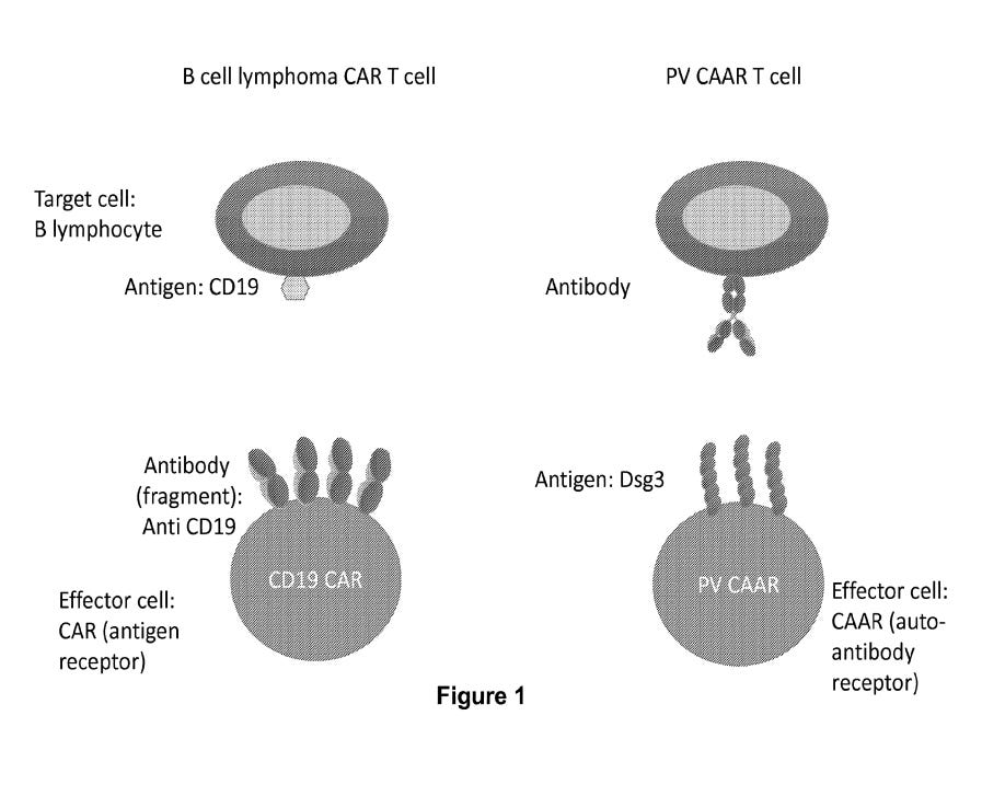
The patent describes a new type of T cell receptor that can be used to target and kill B cells that produce autoantibodies. Autoantibodies are antibodies that attack the body's own tissues. They are a hallmark of many autoimmune diseases, such as pemphigus vulgaris.
The chimeric autoantibody receptor (CAAR) is a fusion protein that combines the extracellular domain of an antibody with the intracellular domain of a T cell receptor. This allows the CAAR to bind to the autoantibody on the surface of a B cell and trigger the T cell to kill the B cell.
One method to produce CAAR-T cells involves genetically engineering T cells to express the CAAR. Another method involves using a virus to deliver the CAAR gene to T cells. The patent also describes several potential applications for the CAAR - treating autoimmune diseases by killing the B cells that produce the autoantibodies. It could also be used to prevent the development of autoimmune diseases by destroying B cells that are susceptible to becoming autoreactive. The CAAR has the potential to be a more targeted and effective therapy than existing treatments for autoimmunity, which often have serious side effects.
https://patentimages.storage.googleapis.com/e9/00/89/78538458ba9e3f/US11578113B2.pdf
How are autoantibodies discovered?
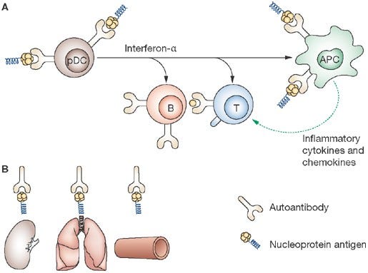
The first autoantibodies were discovered in the late 1940s, when both antinuclear antibodies (ANAs) and rheumatoid factors (RFs) were described. ANAs are antibodies that react with components of the nucleus of cells, while RFs are antibodies that react with the Fc region of immunoglobulins. These autoantibodies are now known to be associated with a variety of autoimmune diseases, including systemic lupus erythematosus (SLE), rheumatoid arthritis (RA), and Sjögren's syndrome. For example, the discovery of the lupus erythematosus (LE) cell was a serendipitous finding. The LE cell is a white blood cell that has engulfed a fragment of nuclear material. This finding was made in the 1940s by researchers who were studying the blood cells of patients with SLE. The LE cell is now known to be a sign of active SLE.
Common methods now to detect autoantibodies are:
- Serological screening: testing blood samples for the presence of autoantibodies against specific self-antigens. This can be done using a variety of techniques, such as enzyme-linked immunosorbent assay (ELISA), immunofluorescence, and Western blotting.
- Cell-based assays: assays involve culturing cells in the presence of autoantibodies to see if they can bind to and damage the cells. This can be a useful way to identify autoantibodies that are pathogenic, meaning that they can cause disease.
- Genetic studies: identify genes that are associated with the production of autoantibodies. This can help to shed light on the underlying causes of autoimmune diseases.
This work is leading to the identification of new autoantibodies that may be useful for diagnosing and treating autoimmune diseases. It has helped us to identify the target antigens of autoantibodies, which has led to the development of new diagnostic tests and treatments for these diseases. Autoantibodies are also being used as biomarkers to track the progression of disease and to predict the risk of relapse.
Scientist Stories: Magdalena Zernicka-Goetz, Human Development and Synthetic Embryos
Zernicka-Goetz is a Professor Caltech & the University of Cambridge
She was first to reveal that cells within the early mouse embryo are not identical but differ in their developmental potential and to apply RNAi to investigate genes directing the fate of individual cells spatially and temporally