
Axial partners with great founders and inventors. We invest in early-stage life sciences companies such as Appia Bio, Seranova Bio, Delix Therapeutics, Simcha Therapeutics, among others often when they are no more than an idea. We are fanatical about helping the rare inventor who is compelled to build their own enduring business. If you or someone you know has a great idea or company in life sciences, Axial would be excited to get to know you and possibly invest in your vision and company . We are excited to be in business with you - email us at info@axialvc.com
Inventors #8
A set of ideas and observations on inventions and discoveries in life sciences.
Immunology
The immune system and everything in it.
Microenvironment mapping via Dexter energy transfer on immune cells - https://science.sciencemag.org/content/367/6482/1091 - the MacMillan Group at Merck invented a tool to label surface proteins around a given target they call MicroMap. They show the power of their tool to map proteins surrounding PD-L1 on B-cells. Through this work, the group identified 6 features that drive the success of cellular mapping tools:
“Catalytic manifold tolerant of aqueous conditions and biomolecules”
A easily-conjugated and modality-agnostic catalyst
Catalyst is activated at a “diffusion-limited rate”
A labeling probe that is only activated within 1 nm (radius) of the catalyst
The probe does not diffuse for too far once activated (i.e. diffusion limited)
The label is agnostic to the catalyst
The group used a catalyst-antibody conjugate in a proof-of-concept study to map surrounding proteins of PD-L1 on B-cells when interacting with T-cells (PD-1/CD45 expressing and finding 11 other targets in PD-L1’s proximity. The MicroMap tool invented here is particularly useful to map microenvironments from immunosynapses to TMEs. Moreover, the tool is useful to map cell-cell interfaces, identify new antibody targets, and more.

Biochemistry and structural biology
The granddaddy of them all.
Targeted Degradation of Transcription Factors by TRAFTACs: Transcription Factor Targeting Chimeras - https://www.biorxiv.org/content/10.1101/2020.10.12.336529v1 - the Crews Lab at Yale developed a set of molecules called TRAnscription Factor TArgeting Chimeras (TRAFTAC) to selectively degrade transcription factors:
TRAFTACs are bifunctional molecules: an oligonucleotide (dsDNA) that binds a given transcription factor is linked to a dCas9 protein fused to a HaloTag. The overall system has another component: a PROTAC fused to a HaloTag to bind the dCas9 in order to engage an E3 ligase to selectively degrade the target.
The group use TRAFTACs on NF-κB and brachyury (relevant for cancer) to show that changes in the oligonucleotide can imbue specificity and lead to targeted degradation
The next step is to use the dCas9 to bind specific sites of the genome to degrade promoter-specific transcription factors. This would be a major step forward because drugging transcription factors is tricky due to the function to control expression of multiple genes and the difficulty to develop very specific small molecules. TRAFTACs have the potential to drug a transcription factor (without needing structural information or a ligand) for a specific gene while not altering expression of other genes controlled by the same transcription factor.

Neuroscience
Roughly 20 years behind but set up to transform the concept of human.
Deep brain optogenetics without intracranial surgery - https://www.nature.com/articles/s41587-020-0679-9 - out of the Deisseroth Lab at Stanford, the group uses the channelrhodopsin ChRmine to activate neural circuits in mice brain up to 7 mm in depth:
ChRmine is a channelrhodopsin that is red-shifted light sensitive (100x improved versus comparable opsins) and is extremely useful for two-photon imaging studies due to its fast off-kinetics. It was discovered through mining various microbial genomes.
With these advantages, the group decided to use ChRmine to do a PoC study of “implant-free deep brain optogenetics”
The group used viral delivery (AAV8) of ChRmine to get to neurons within the ventral tegmental area (VTA), which is 4.5 mm from the skull. They tested the upper limits of their system, they did injections at different depths in a rat model; they found a signal at 7 mm from the skull surface. From this work, the group did not find substantial tissue damage in the rat models used.
This PoC study is exciting because it shows that ChRmine can be used to activate specific neurons in deeper parts of a brain in a model without surgery.

Cell biology
Cell structure and function.
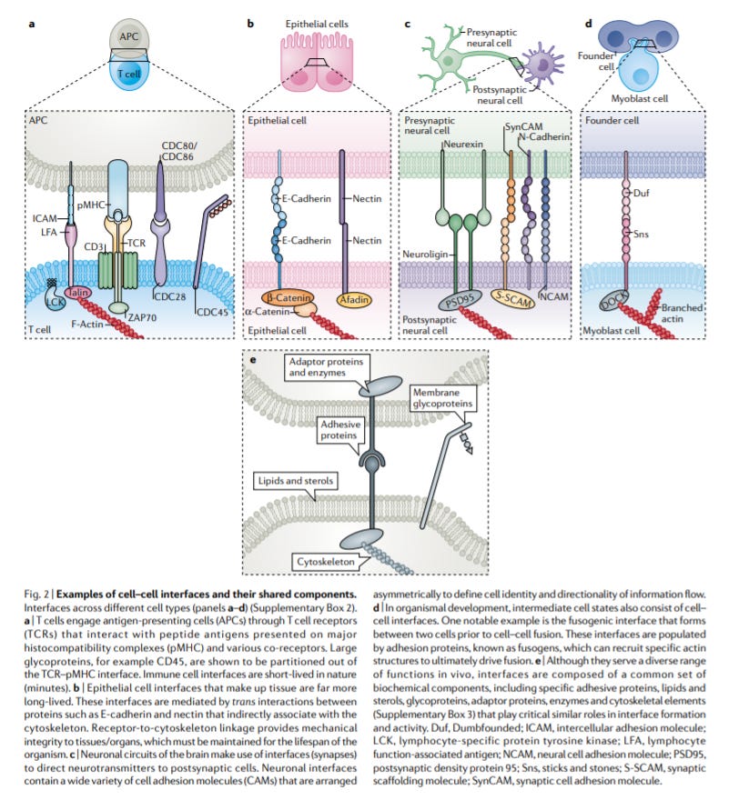
Cell–cell interfaces as specialized compartments directing cell function - https://www.nature.com/articles/s41580-020-00298-7?WT.mc_id=TWT_NatRevMCB - the Fletcher Lab at UC Berkeley put out a very useful review on the biology of cell-cell interfaces. His lab is one of the world leaders in mechanobiology:
Cell–cell interfaces are important for an individual cell to respond to its environment. From the origins of multicellular life to neurons and immune cells, the interaction between cells drives development and disease.
Various tools from microscopy, optical tweezers, MicroMap, and other sensors have been improving to enable the study of these interfaces
These interfaces have a few features that make them unique and valuable to study/engineer: (1) compartments of constrained targets (2) enable cells to respond quickly to a new input (3) these two parts create new therapeutic avenues especially for undrugged targets (this theme also applies to condensates)
Cell-cell interfaces can be temporally short (i.e. immune cells) to long (i.e. neurons) and also cover large surface areas (i.e. postsynaptic membranes between neurons) to small ones (i.e. epithelial cells)
The review suggests a few therapeutic pathways for immunotherapy: (1) targeting interfaces directly if they driven disease pathology (2) shorten cell-cell interfaces to maximize the efficacy of CAR-T cell therapies (3) inventing mechanosensitive therapies that may be distinguishing between diseased and healthy cells
From the review a few outstanding questions in the field are: “How does the diversity of cell surface biomolecular size and the extent of their post-translational modifications shape interfaces and influence function? Is it possible to engineer cell–cell interfaces in vivo to modulate cell function for a desired effect or even to create new cell functionalities with novel, synthetic interfaces?”
Genetics, genomics, and developmental biology
Heredity and variation.
Topological constraints in early multicellularity favor reproductive division of labor - https://elifesciences.org/articles/54348 - the Yunker Lab at Georgia Tech developed a model to connect the topology of early multicellular life to reproductive specialization. Conventional wisdom in the field is that multicellularity only evolved if the return on investment (ROI) for specialization is higher than generalism. The Yunker Lab created a model to mimic the potential early topology of life by restricting cells ability to only interact and share resources with neighboring cells. With this model, the group found that specialization can still occur even if the ROI is negative because the incentives to reproduce the multicellular structure/topology is greater than the individual cell. This leads to soma cells giving more resources to reproductive cells that can be shown to be selfish by the latter. Overall, the work does a great job to establish that multicellular life had a lot more pathways to emerge. The King Lab at Berkeley has done pioneering work on the origins of multicellularity. They use choanoflagellates as a model organism.
